Bone healing.
Skeletal fracture occurs when bone absorbs sufficient energy under mechanical loading to fail, resulting in cortical discontinuity.
Fracture healing is the biological process by which trabicular discontinuity of a bone is restored.
This requires temporal coordinated action of several different cell types, proteins and the expression of hundreds of genes working towards restoring its structural integrity without scar formation.Fracture healing is nothing but reiterating embryonic endochondral bone formation
Fracture healing is unique.
- Resulting bone both microscopically and macroscopically completely resembles that present prior to injury.
- Bone repair is similar to those observed during embryonic bone formation and this is therefore truly a regenerative process.
- The essential difference between bone healing and the healing of any other tissue is unlike any other tissue, bone heals with out scar.
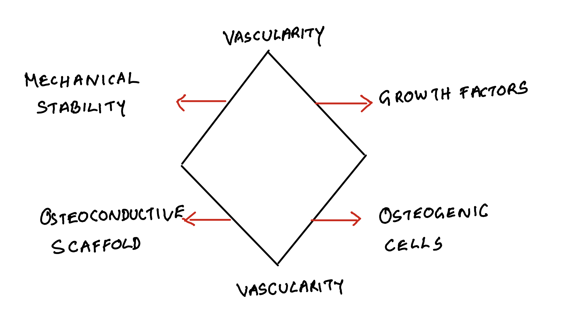
Diamond Concept
Cortical and cancellous bone heal by slightly different mechanisms. Interplay between at least four different factors are identified which can greatly effect this process.
- Stability
- The scaffold
- Growth factors
- Osteo-progenitor cells.
This is the basis of Diamond concept of healing which we will discuss later.
Perrin’s strain theory
When given a chance to heal with minimal gap in between the fractured ends, bone heals by primary intention which is called primary healing. In other situations with bigger gaps in between the fractured ends, they heal by secondary intention, which is called secondary healing. This effect of gap on the fracture healing process is due to its affect on the strain which is called fracture gap strain which is given by the formula ∆FGap/FGap as described by Perrin.
Strain is the deformation of a material when a given force is applied.
Fracture gap stain is the ratio of relative displacement of fracture ends versus initial fracture gap width.
Percentage of strain determines the type of tissue formed during the healing. The strain will be less-
- With large fracture area (less ∆FGap)
- More number of fracture fragments.
- Less gap in between the fracture fragments.
Strain and bone healing
| 100 |
Granulation |
* |
Absolute instability |
| 17 |
Fibrous tissue |
* |
Unstable |
| 2-10 |
Fibrocartilage |
20 healing |
Relative stability |
| <2 |
Lamellar bone |
10 healing |
Absolute stability |
Percentage of strain determines the type of tissue formed during the healing.
Primary bone healing.
Cortical bone
Cortical bone heals by Osteonal remodeling by Osteonal cutting cones if there is no gap. This requires
- absolute stability (with strain < 2%)
- No gap
- Viable bone ends
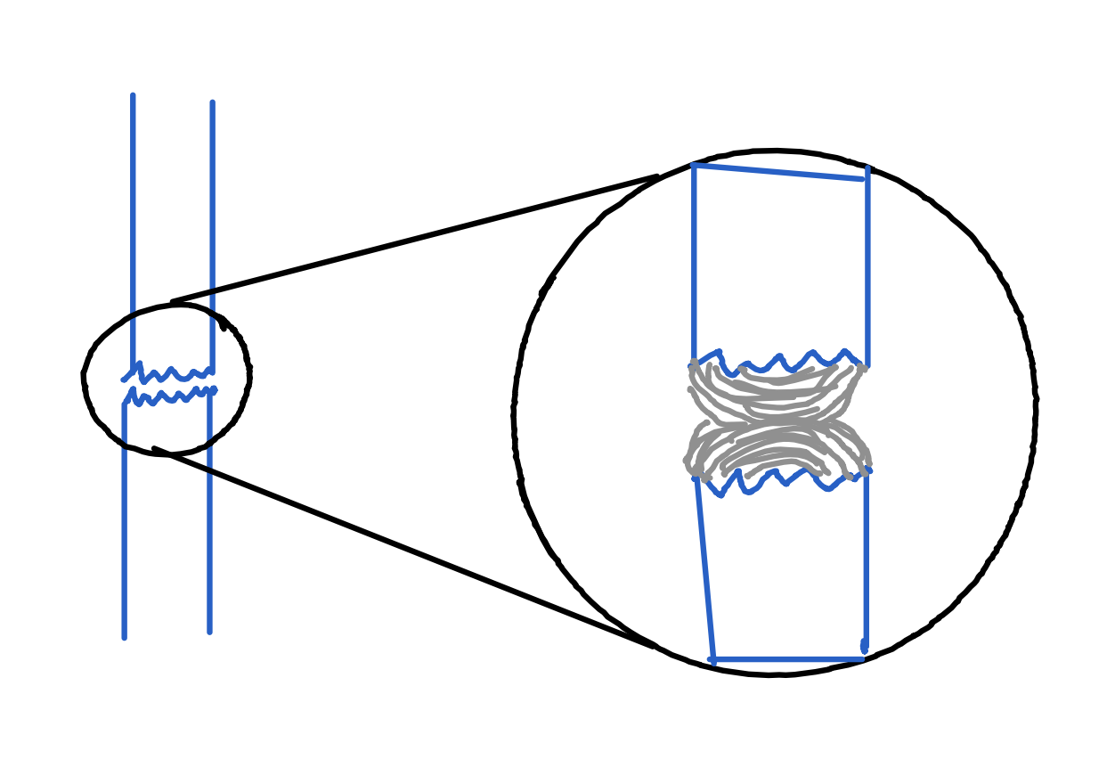
If there is gap but < 500µm, healing is by gap healing. In gap healing, osteoblast lays down sequential layers of bone to close the gap.
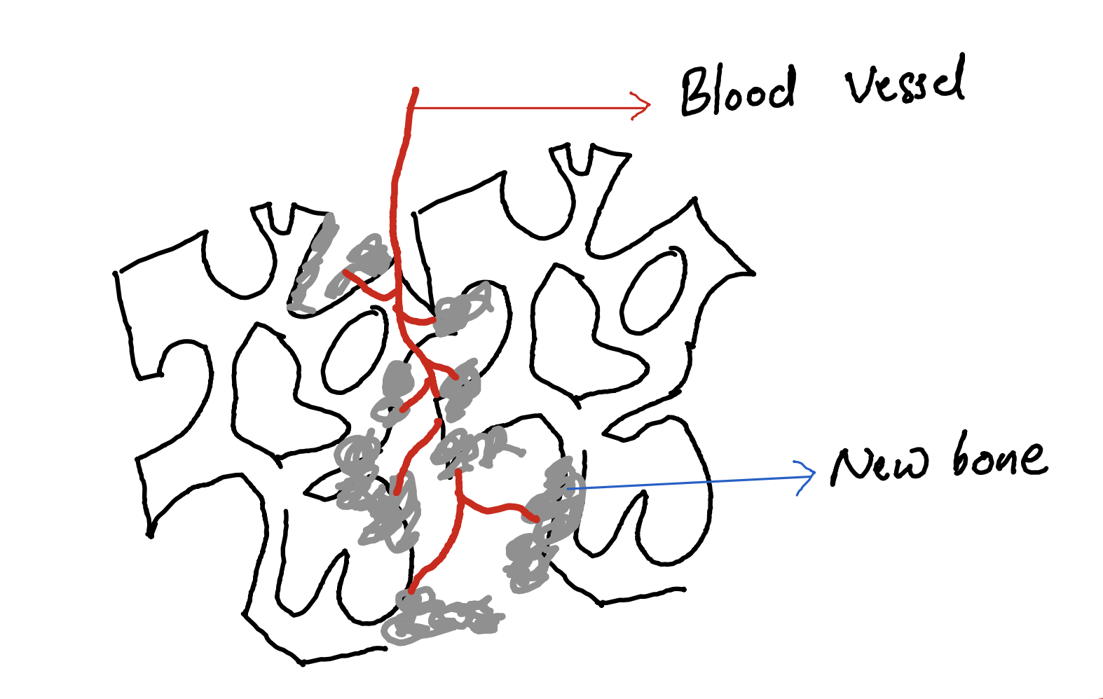
Cancellous bone.
Heals primarily by a mechanism called bony ingrowth. Here revascularisation followed by osteoblasts laying down new bone.
Secondary bone healing
Ham in 1930 published a description of early phases of bone healing after studying fractured rabbit fibulae and ribs. Phases of bone healing was more fully described by McKibbin in 1978. In simple terms, bone heals in 3 phases, like any other tissue.
Phases of bone healing
- Inflammation
- Cellular proliferation and differentiation
- Remodeling.
However the process is complicated by high specialisation and calcified nature of bone.
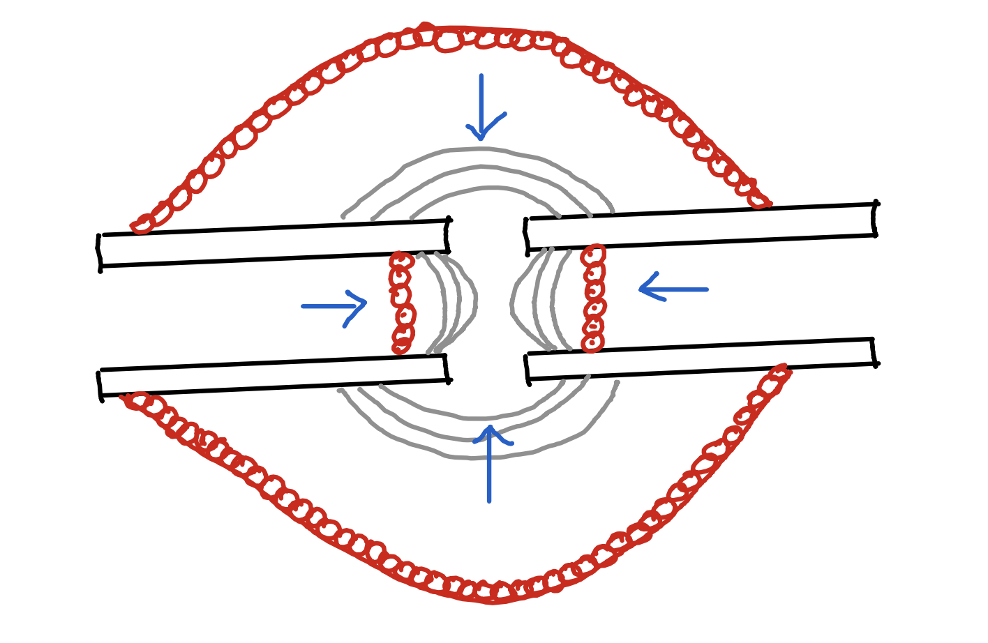
Differentiation of primitive mesenchymal stem cells in to cells with osteogenic potential result in a specialized connective tissue formation called callus. Ambient strain stimulate fracture gaps to form callus . For this the strain environment should be 2%-10%. Bio-mechanical function of the callus is to reduce strain by
- increasing the diameter of the fractured area.
- Increasing the stiffness.
Soft and Hard callus.
Soft callus: Central, due to endochondral ossification.
Hard callus: Peripheral, due to intramembranous ossification
Remodelling
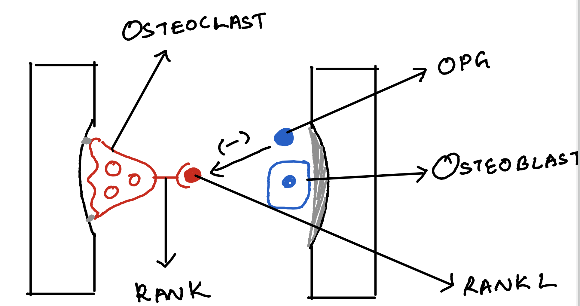
20 process directed by osteoclastic resorption by howship lacunae in metaphysis through Osteons in the cortices. Involves complex interplay of RANK-RANKL
Back to top